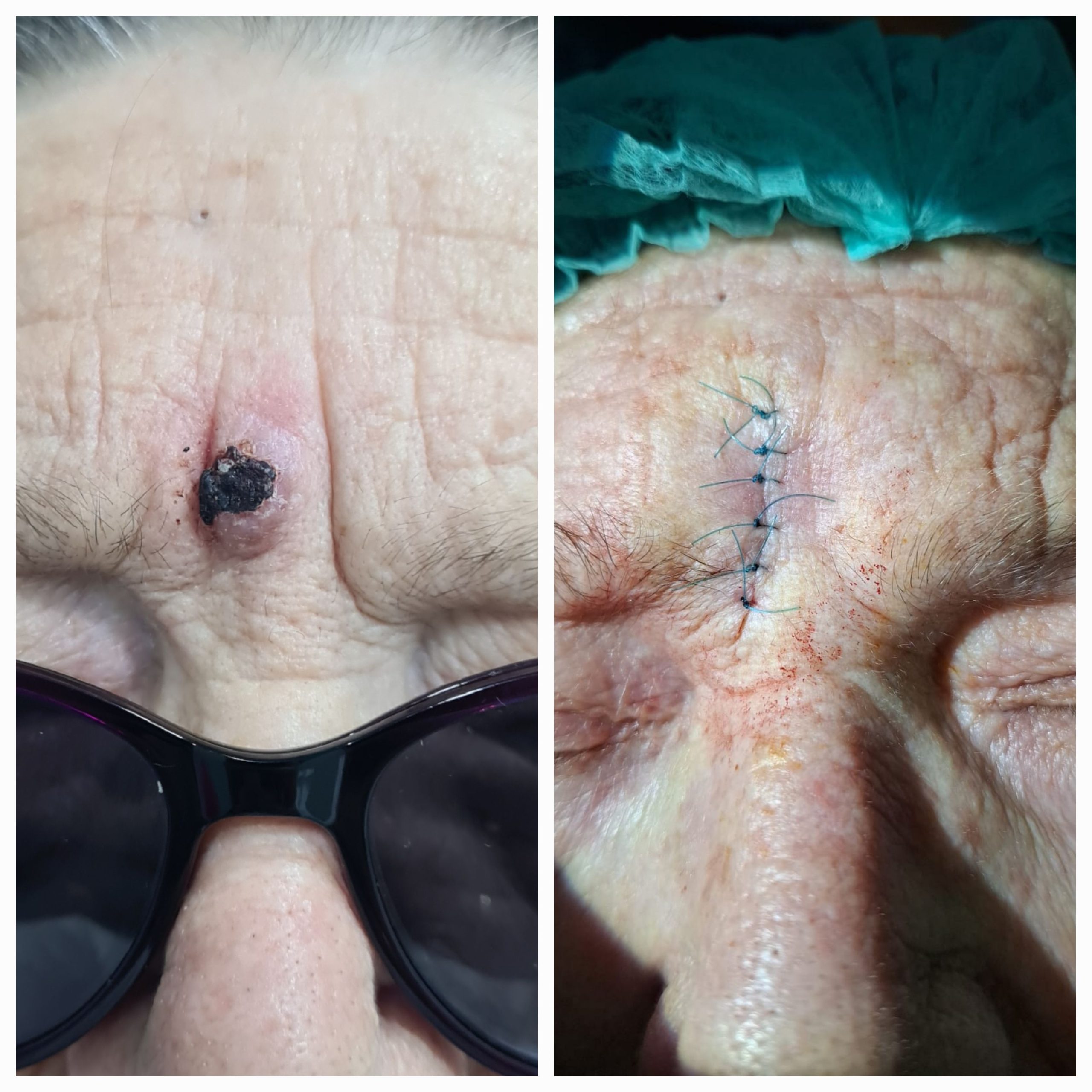
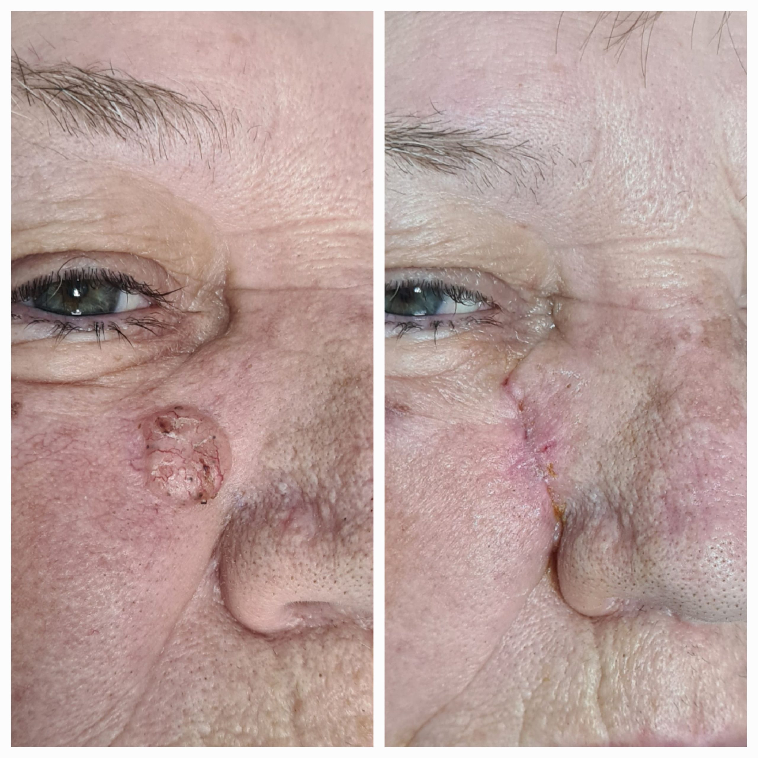
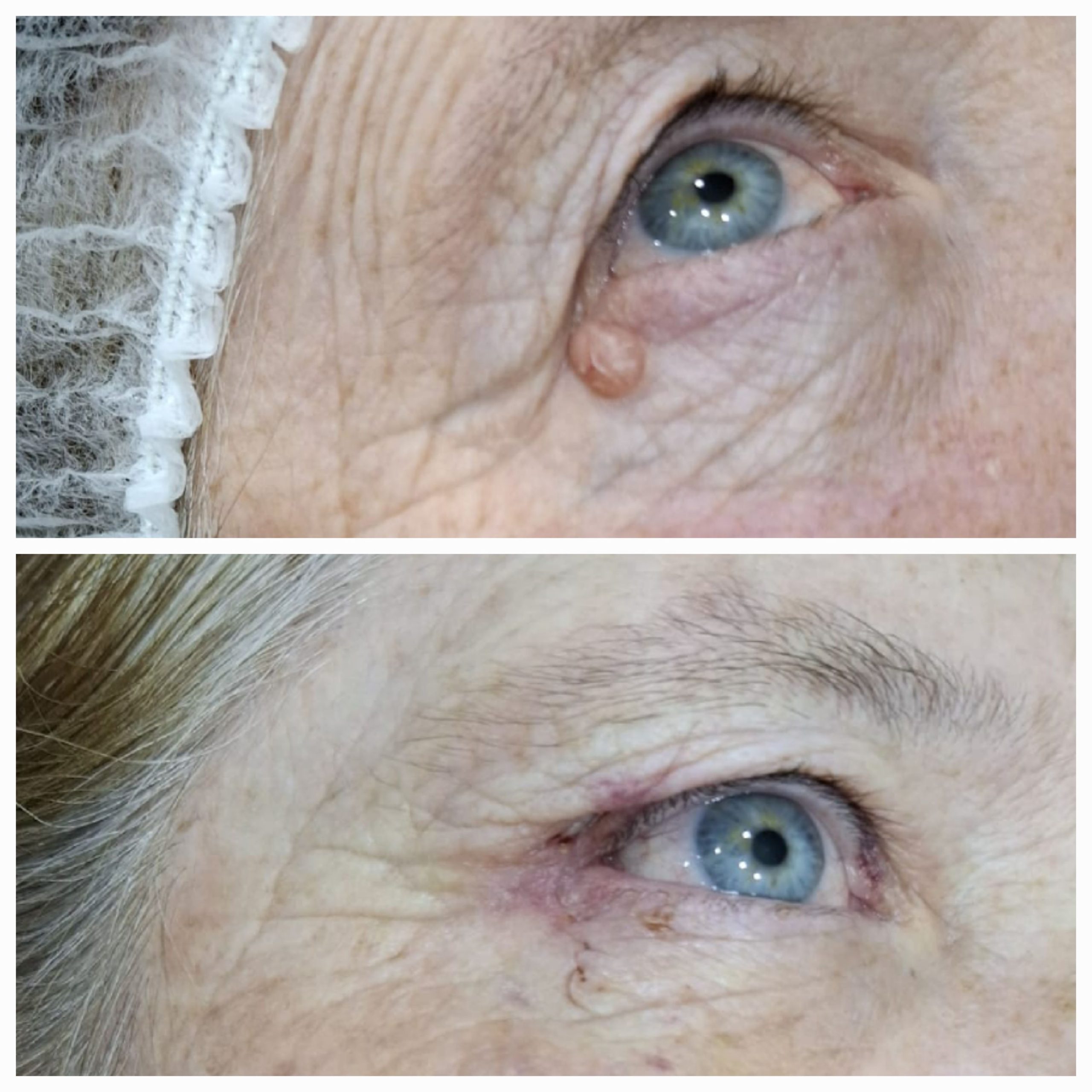
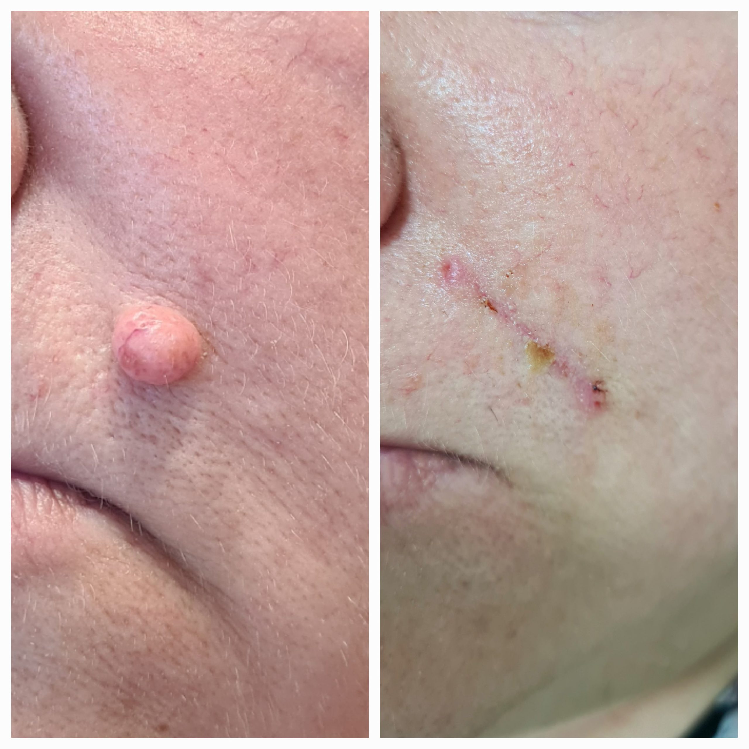
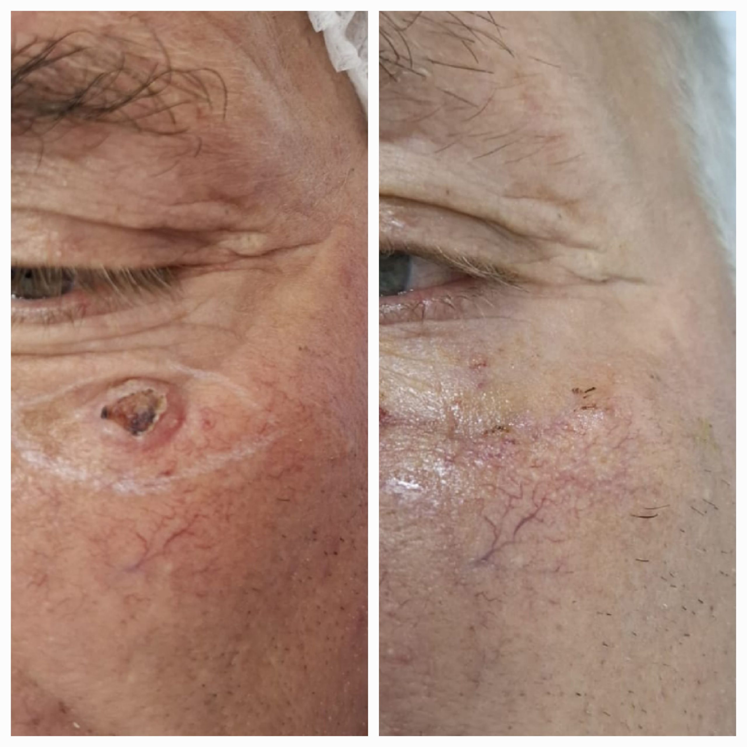
The surgical treatment aims to excise the tumor within oncological safety margins.
After tumor removal, the area is aesthetically reconstructed to restore shape and function.
Depending on the type, size of the lesion, and complexity of the intervention, defect coverage with a skin graft or flap may be necessary.
The duration of the procedure varies between 45 and 60 minutes.
Patient preparation takes between 15 and 20 minutes.
Dr. Simion Răzvan has extensive experience in the surgical removal of tumoral formations, having successfully performed over 300 interventions.
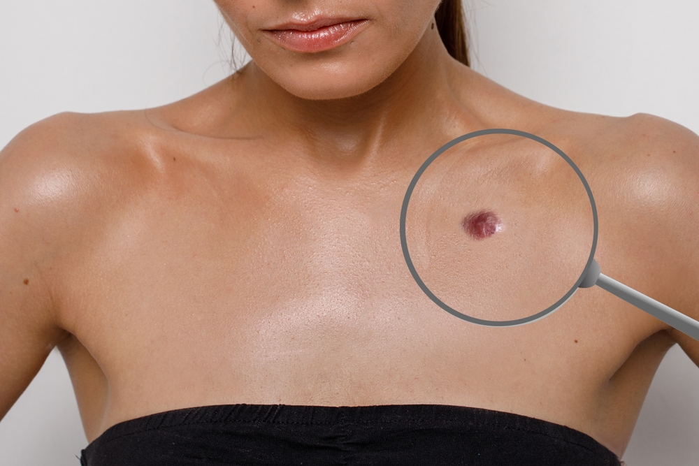
Tumoral formations can be benign (non-cancerous) or malignant (cancerous).
Depending on their location, they can be cutaneous (on the skin) or subcutaneous (under the skin), varying in shape and size from a few millimeters to several centimeters.
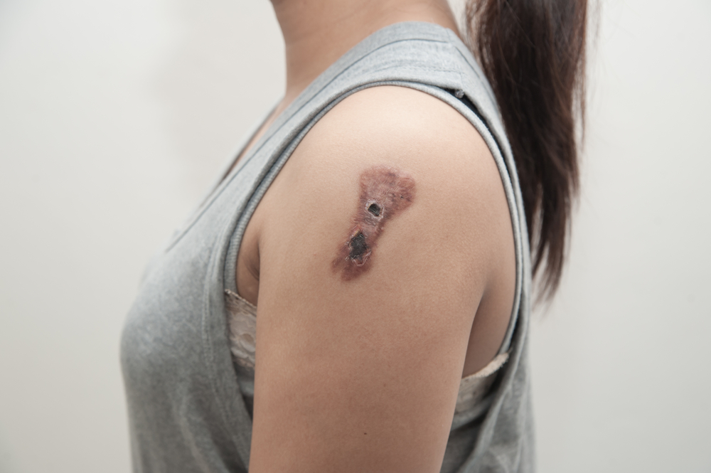
If they grow in size and are neglected, they can ulcerate, bleed, and become infected.
Therefore, it is important that they are diagnosed and excised at an early stage.
Depending on the size of the tumor, the remaining defect can be closed either with sutures or, in the case of larger defects, with a covering graft.
After excision, it is recommended that the tumoral formation be sent to the laboratory for histopathological examination to obtain a definitive diagnosis.
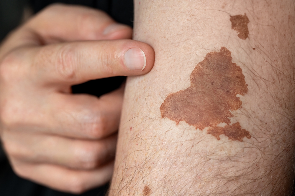
Melanoma (skin cancer) appears in various shapes, sizes, and colors, making it difficult for patients to recognize this type of tumor. It is important to remember the ABCDE mnemonic for detection:
Asymmetry of the tumor
Borders that are irregular or poorly defined
Color with shades of brown, black, gray, red, or white
Diameter larger than 6 millimeters
Evolution – the lesion is new or has changed in size, shape, or color.
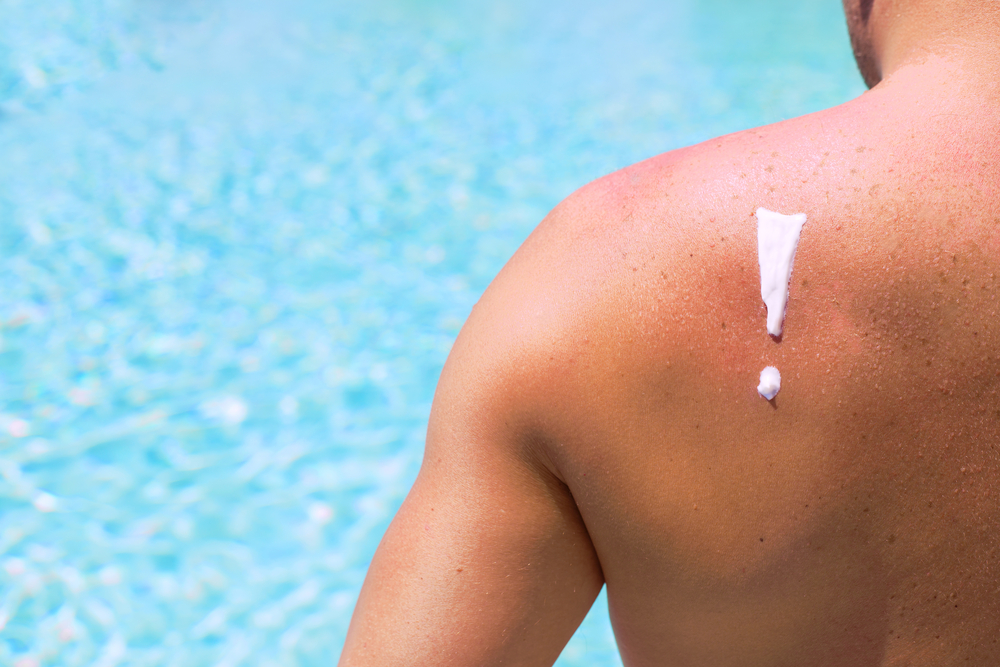
Men are more prone to developing melanoma on the body surface, often on the back, while in women it more frequently appears on the face or legs.
Since early detection is vital, immediate surgical removal is recommended. After excision within oncological safety margins, histopathological examination and immunohistochemical tests are advised.